
Epithelial-Mesenchymal Transition and Fibrosis Markers in the Epitalon Retinal In Vitro Study: Technical Insights and Evidence Limitations
This resource examines the meaning and significance of epithelial-mesenchymal transition (EMT) and fibrosis-associated markers in the context of an in vitro study investigating Epitalon’s effects on human retinal pigment epithelial cells under high-glucose conditions. It details the experimental findings, the roles of EMT and fibrosis in diabetic retinopathy models, and the limitations of translating these results to clinical contexts.

Introduction: Context of EMT and Fibrosis in Retinal Research
Epithelial-mesenchymal transition (EMT) and fibrosis are central processes in the pathogenesis of proliferative diabetic retinopathy (PDR) and other retinal diseases. EMT describes a cellular program where epithelial cells lose their polarity and adhesion, acquiring mesenchymal, migratory, and fibrotic properties. In the retina, this transition is implicated in subretinal fibrosis, a hallmark of advanced diabetic eye disease. Fibrosis-associated markers, such as vimentin, fibronectin, and alpha-smooth muscle actin (α-SMA), reflect the activation of this process and the deposition of extracellular matrix components that can disrupt retinal architecture and function.
A recent in vitro study investigated the synthetic tetrapeptide Epitalon (Ala-Glu-Asp-Gly) in a model of diabetic retinopathy using high-glucose (HG)-injured human retinal pigment epithelial (ARPE-19) cells. The study focused on how Epitalon modulates EMT and fibrosis markers under hyperglycemic stress, aiming to elucidate potential mechanisms relevant to delayed wound healing in diabetic retinal disease Source 1.
Defining EMT and Fibrosis-Associated Markers
Epithelial-Mesenchymal Transition (EMT)
EMT is a biological process where epithelial cells lose their cell-cell adhesion and polarity, transitioning into a mesenchymal phenotype characterized by increased motility and invasiveness. In the context of retinal pigment epithelial (RPE) cells, EMT is triggered by pathological stimuli such as chronic hyperglycemia, leading to the loss of epithelial characteristics and the acquisition of fibrotic, migratory properties. This process is implicated in the formation of fibrotic membranes and subretinal scarring observed in proliferative diabetic retinopathy and other retinal disorders.
Fibrosis-Associated Markers
Fibrosis markers are molecular indicators of the transition from normal tissue repair to pathological scarring. In the study, the following markers were assessed: - Vimentin (VIM): An intermediate filament protein upregulated during EMT, indicating a shift toward a mesenchymal phenotype. - Fibronectin (FN1): An extracellular matrix glycoprotein involved in tissue repair and fibrosis. - Alpha-smooth muscle actin (ACTA2, α-SMA): A cytoskeletal protein characteristic of myofibroblasts, cells central to fibrotic tissue remodeling.
Transcription factors such as SNAIL1, ZEB1, and TWIST1 regulate EMT by repressing epithelial genes and activating mesenchymal gene expression. Their upregulation is a hallmark of EMT activation Source 1.
Experimental Model and Methods
The referenced study used the ARPE-19 human retinal pigment epithelial cell line, a widely accepted in vitro model for retinal research. Cells were exposed to high-glucose (35 mM) conditions to mimic the hyperglycemic environment of diabetic retinopathy. The experimental design included: - Scratch wound assay: To assess wound healing and cell migration. - Quantitative PCR (qPCR): For mRNA expression of EMT and fibrosis-related genes (SNAIL1, ZEB1, TWIST1, VIM, FN1, ACTA2). - Western blot: For protein expression of α-SMA. - Reactive oxygen species (ROS) assays: To measure oxidative stress.
Epitalon was tested at concentrations of 20, 40, and 60 ng/mL. Mannitol was used as an osmotic control to rule out non-specific effects of increased medium osmolarity Source 1.
Key Findings: EMT and Fibrosis Markers in High Glucose and Epitalon Conditions
Effects of High Glucose
Exposure of ARPE-19 cells to high glucose resulted in: - Delayed wound healing: Slower closure of scratch wounds compared to standard glucose conditions. - Increased ROS production: Elevated intracellular hydrogen peroxide, indicating oxidative stress. - Downregulation of antioxidant genes: Reduced expression of SOD2, CAT, and HMOX1. - Upregulation of EMT transcription factors: Significant increases in SNAIL1, ZEB1, and TWIST1 mRNA. - Increased fibrosis markers: Elevated mRNA levels of VIM, FN1, and ACTA2, and increased α-SMA protein expression.
These changes collectively indicate that high-glucose conditions promote EMT and fibrotic transformation in RPE cells, consistent with the pathophysiology of diabetic retinopathy Source 1.
Effects of Epitalon
Epitalon treatment in HG-injured ARPE-19 cells led to: - Restoration of wound healing: Improved scratch closure rates at all tested concentrations. - Reduction in ROS: Lowered hydrogen peroxide levels, suggesting antioxidant activity. - Normalization of antioxidant gene expression: Partial restoration of SOD2, CAT, and HMOX1 mRNA levels. - Suppression of EMT transcription factors: Dose-dependent reduction in SNAIL1, ZEB1, and TWIST1 expression. - Inhibition of fibrosis markers: Decreased VIM, FN1, and ACTA2 mRNA, and reduced α-SMA protein levels.
These findings suggest that Epitalon can counteract hyperglycemia-induced EMT and fibrotic gene expression in this in vitro model Source 1.
Biological Significance of EMT and Fibrosis Markers in Retinal Disease Models
EMT in Retinal Pathology
EMT in RPE cells is a critical event in the development of subretinal fibrosis and proliferative membrane formation, which can lead to retinal detachment and vision loss in advanced diabetic retinopathy. The upregulation of EMT transcription factors (SNAIL1, ZEB1, TWIST1) and mesenchymal markers (vimentin, fibronectin, α-SMA) reflects a shift toward a fibrotic, migratory phenotype that disrupts normal retinal structure and function.
Fibrosis Markers as Indicators of Pathological Remodeling
The increased expression of vimentin, fibronectin, and α-SMA indicates activation of myofibroblast-like cells and excessive extracellular matrix deposition. In the context of diabetic retinopathy, this process underlies the formation of fibrotic membranes and scarring, contributing to irreversible retinal damage Source 1.
Mechanistic Insights: How Epitalon May Influence EMT and Fibrosis in Vitro
The study proposes several mechanisms by which Epitalon may modulate EMT and fibrosis markers: - Antioxidant activity: By reducing ROS levels, Epitalon may alleviate oxidative stress, a known trigger of EMT and fibrotic signaling. - Epigenetic modulation: The study observed that Epitalon prevented HG-induced global DNA hypomethylation, suggesting a potential role in regulating gene expression at the epigenetic level. - Transcriptional regulation: Suppression of EMT transcription factors and fibrosis-associated genes points to a direct or indirect effect on signaling pathways involved in cellular plasticity and extracellular matrix remodeling.
However, the precise molecular targets and signaling cascades affected by Epitalon remain to be fully elucidated Source 1.
Study Limitations and Considerations
In Vitro Model Constraints
- Cell line limitations: ARPE-19 cells, while widely used, may not fully recapitulate the complexity of native human RPE physiology.
- Glucose concentrations: The standard culture medium for ARPE-19 already contains higher-than-physiological glucose levels, and further elevation to mimic hyperglycemia may not perfectly model in vivo diabetic conditions.
- Lack of systemic factors: The in vitro system does not account for the influence of circulating mediators, immune cells, or chronic metabolic changes present in diabetic patients.
Translational Gaps
- No in vivo or clinical data: The findings are limited to cell culture and cannot be extrapolated to animal models or human outcomes without further research.
- Unresolved mechanisms: While the study demonstrates changes in gene and protein expression, the downstream functional consequences and long-term effects remain unclear.
- No safety or efficacy conclusions: The study does not address the safety, pharmacokinetics, or therapeutic potential of Epitalon in humans or animals.
Unresolved Questions and Future Research Directions
- Mechanistic pathways: Further studies are needed to clarify how Epitalon modulates EMT and fibrosis at the molecular level, including potential interactions with signaling pathways such as Akt2 and ERK.
- Epigenetic effects: The impact of Epitalon on DNA methylation and its relevance to gene regulation in diabetic retinopathy warrants deeper investigation.
- Advanced models: Use of three-dimensional cell cultures, organ-on-chip systems, and animal models could provide more physiologically relevant insights.
- Clinical translation: Rigorous preclinical and clinical studies are required to assess the safety, dosing, and efficacy of Epitalon for retinal diseases.
Key Takeaways
- EMT and fibrosis markers in the referenced study serve as indicators of pathological cellular transformation and tissue remodeling in a diabetic retinopathy model.
- Epitalon was shown in vitro to suppress hyperglycemia-induced EMT and fibrosis-associated gene and protein expression, potentially via antioxidant and epigenetic mechanisms.
- All findings are limited to an in vitro cell culture model and do not imply clinical efficacy, safety, or regulatory approval for Epitalon in any human or veterinary context.
- Further research is necessary to validate these results in more complex biological systems and to elucidate the underlying mechanisms.
For research and educational purposes only. Not medical advice. Research products are not for human or veterinary use.
Sources
- The Antioxidant Tetrapeptide Epitalon Enhances Delayed Wound Healing in an in Vitro Model of Diabetic Retinopathy - PMC. https://pmc.ncbi.nlm.nih.gov/articles/PMC12356729/
Continue exploring
TRACE THE EVIDENCE
Sources & further reading
RELATED PRODUCT INFORMATION
From the Certified Pep catalog.
Product references are informational, not a recommendation for human use. Prices and availability must be verified at retailer checkout.
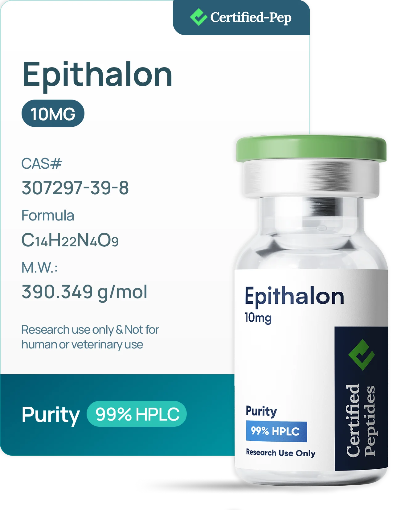
EPITHALON 10MG
USD 54.00 USD 45.00
In stock
Last synced: 2026-11-01 10:07
Add to retailer cart ↗View product details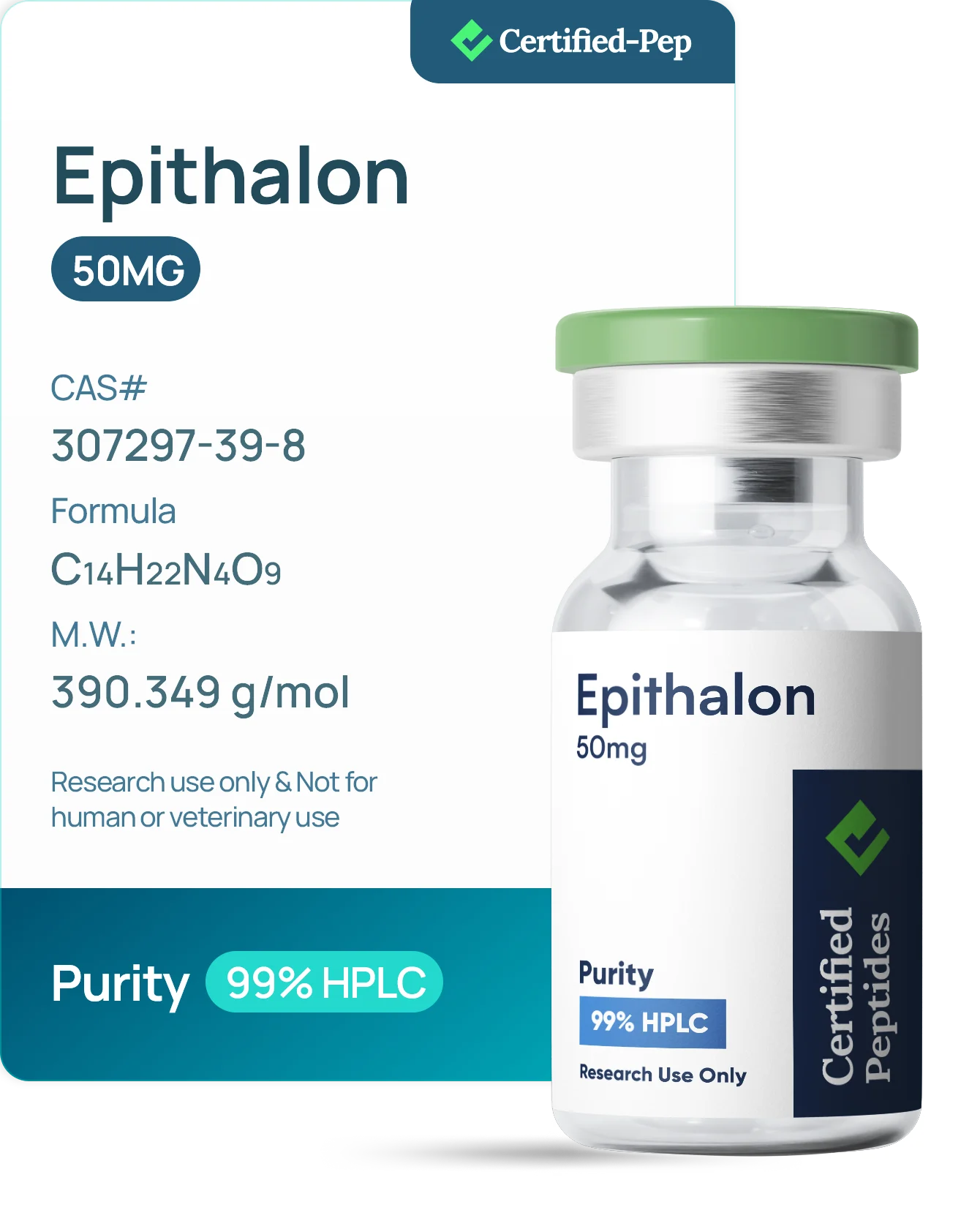
EPITHALON 50MG
USD 134.00 USD 110.00
In stock
Last synced: 2026-11-01 10:07
Add to retailer cart ↗View product details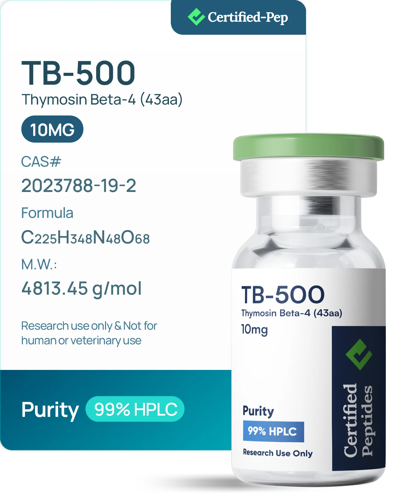
TB-500 10 MG
USD 70.00
In stock
Last synced: 2026-11-01 10:07
Add to retailer cart ↗View product details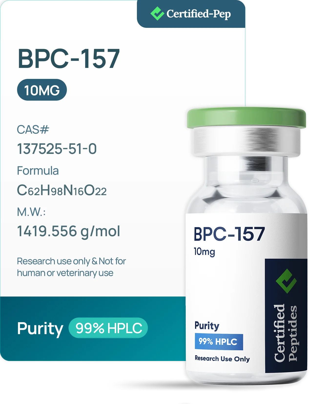
BPC-157 10 MG
USD 60.00
In stock
Last synced: 2026-11-01 10:07
Add to retailer cart ↗View product details
BPC-157 20 MG
USD 90.00
In stock
Last synced: 2026-11-01 10:07
Add to retailer cart ↗View product details
BPC-157 500 mcg 60 CAPSULES
USD 160.00 USD 100.00
In stock
Last synced: 2026-11-01 10:07
Add to retailer cart ↗View product details